Behavioral Neuroscience I
What is the Brain made of?
Neuroanatomy: Anatomy of the Brain
Things to Remember:
- The brain is super complicated with a laundry list of parts + regions
- Scientists like to make up fancy words so they sound smart
- Anatomy, especially neuroanatomy, is about learning these made-up words so you can talk about very specific areas of the body.
In short: There is going to be a lot of vocab in this section. I’m sorry.
Unless you have a test on this subject, I don’t recommend memorizing anything. Getting the general idea of how things work is much more important than remembering the difference between the Superior Colliculus and Inferior Colliculus.
I will also try my best to cut out as much medical jargon as possible. I think it distracts from understanding the fundamental concept of neuroanatomy, which is:
How is the brain structured?`
We are going to start as general as possible and quickly work our way down to neurons, the building blocks of the brain.
Brain 101
Parts of the Nervous System
Like it sounds, the Nervous System is a collection of connected nerves that helps the body communicate with other body parts. The brain is central to this communication. The Nervous System is made of:
- Central Nervous System: Anywhere in the body with nerves that are encased in bone:
- Brain
- Spinal cord
- Peripheral Nervous System: Anywhere in the body with nerves that are not encased in bone:
- Nerves leaving brain or spinal cord
- Nerves connected to internal organs
Brain Material
The brain itself is made up of:

- grey matter: Neuron synapses (connections) and cell bodies found along the “outside” of brain
- white matter: Mylinated (fat-covered) cells found “inside” the brain
The brain floats in a liquid called Cerebral Spinal Fluid (CSF), which acts as a shock absorber.
The brain is protected by bone and the meninges, membranes covered in pain receptors. This is where the word meningitis comes from, and why meningitis hurts so bad.
There are also 4 pockets of CSF fluid “within” the brain called ventricles.
Brain Lobes & Cortices
There are four brain lobes, named for the bones overlying the skull. The names are quite poor and don’t actually reflect their function, but roughly:
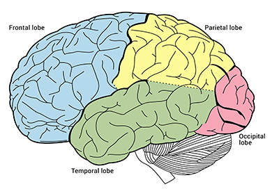
- Occipital Lobe: Back of brain. Home of visual system
- Temporal Lobe: Left side of brain. Home of auditory system
- Parietal Lobe: Top + Right side of brain. Home of body representation and movement
- Frontal Lobe: Front of brain. Home of planning and higher processing
While the brain lobes are quite popular, they are not the most accurate way to understand brain areas. A better representation can be seen in the sensory cortices:

As you can see from the above image, some brain regions have helpful names like Visual Cortex and some are named after scientists who really wanted to be famous like Broca’s Area. We will breakdown these cortices during our Tour of the Nervous System.
Neuron 101

Neurons are made up of 4 parts:
- Dendrites, which receive neuron inputs
- Cell Body, which integrates all inputs
- Axons, which transports signal to neuron endings
- Synaptic Terminal, which outputs signal
When talking about neurons, it is important to keep in mind the scale of things– specifically how unbelievably small they are.
- Neuron cell bodies are 10 microns in length, or 1/100th of a millimeter
- Synapses are 1 micron in length, or 1/1000th of a millimeter
- Synaptic Clefts are 10 nanometers, or 1/1000th of a nanometer
For a better visual- the head of a pin is 1.5mm. On a single pin head we could fit…
- 150 neurons cell bodies
- 1,500 synapses
- 1,500,000 synaptic clefts
We have 100 billion neurons in our brain.
To put this in perspective– 100 billion seconds is roughly 3,170 years.
If laid down in a straight line, our neurons, despite being so small you can fit 150 on the head of a pin, would stretch 1000km (621 miles).
Neurons are minuscule in size, and we have an astronomical amount of them.
The Axon
Axons are the roadways between neuron inputs and outputs. While the dendrites, cell body, and synaptic terminal stay relatively the same size, Axons can extend to pretty huge lengths. Like across the entire brain. Or up the entire spinal cord.
Transporting signals this long of distance is hard, and signals can easily get lost or take too long to reach their destination.
To ensure signals are not lost, axons are covered in myelin, or a layer of protective fat.
To ensure the signal can travel fast enough, the layer of myelin (myelin sheath) is broken up with exposed pieces of axon. The “exposures” are called Nodes of Ranvier.
Electrical signals can “jump” between Nodes of Ranvier instead of travel up the entire axon, helping them travel much faster.
The Synapse

Synapses are the endings of synaptic terminals, and are how signals are passed between neurons. The space between neurons is called the synaptic cleft. Tiny bubbles called synaptic vesicles dump even tinier chemicals called neurotransmitters into this cleft, which are then absorbed by receptors on the neighboring neuron (dendrite).
The communication between neuron is fundamental to neuroscience and we will revisit the synapse soon, this was just to familiarize yourself the anatomy/vocab.
Transport & Cleanup
To stay clean, neurons have to both transport new cell parts up to the synaptic terminals (Anterograde Transport) and cleanup old cell parts from axon (Retrograde Transport)

Glial Cells, amongst other things, are specialty cells that help with cleaning and transport. There are four types:
- Astrocytes: Maintain neuron homeostasis by supplying/taking away neurotransmitters.
- Oligodenricytes: Produce myelin for cells in Central Nervous System
- Microglia: Immune system of the brain. Grow large (macrophages) and “eat” foreign bodies
- Schwann Cells: Produce myelin for cells in Peripheral Nervous System
Tour of the Nervous System
Everything has to do with your nervous system -Jonathon Lipnicki
The fundamental concept of this section is:
How does the Nervous System work?
We are going to structure our tour of the nervous system by separating neuroanatomy into
- Input: How signals are first generated
- Processing: How signals are interpreted
- Output: How the body responds to signals
Input
Most signals enter the nervous system through nerves (duh) which are a collection of axons that carry information to and from the CNS.
There are 12 cranial nerves which carry information about senses in the head and internal organs.
Spinal Nerves carrying sensory information enter the CNS through the Dorsal Root, which is a fancy word for the back of the spine. Motor information exits the CNS through the Ventral Root, or front of spine
All sensory inputs (except smell) then travel to the Thalamus, the post office of the brain, to be distributed to appropriate brain region.
Processing
+Pg+440.jpg)
As the above picture shows, there are a lot of brain regions. If you want to memorize all of them, eat your heart out.
Otherwise, here are a few important areas I’ve chosen to highlight:
Cerebral Cortex
The “wrinkly” part of the brain that wraps around the outside. It is essentially a 2-D sheet, with a depth of only about 4mm.
It is responsible for perception, learning, memory, planning, and decisions. Also 40% of the cortex is dedicated to vision.
Cerebellum
The back-bottom of the brain that looks separate from the wrinkly cerebral cortex.
It is responsible for reconciling sensory and motor maps, synaptic plasticity, and motor coordination.
Brainstem
Comprised of the midbrain, Pons, and Medulla. Often called the “Reptilian brain” because it is older and deals with more fundamental processes.
It is responsible for breathing, heart rate and pressure, eating, and sexual arousal.
Output
The literal output of the nervous system comes in the form of a memories, hormones, and behaviors. While later posts will deal with these outputs more in depth, right now we will briefly go over a few key structures:
Limbic System
Primary Structures are:
- Hippocampus: Deals with transferring short-term memory to long-term (not where memories are stored however). I’m sure you’ve noticed that attention, smell, and emotion have a particularly strong effect on memories. This is because the structures responsible for those attributes (Cingulate Gyrus,Olfactory Bulb,and Amygdala respectively) are closely wired to the Hippocampus.
- Hypothalamus: Regulates hormone levels in the body. Also controls the Autonomic Nervous System
Autonomic Nervous System
An automatic response system that spans both the CNS and PNS. It is broken down into two parts:
- Sympathetic Division: The “fight or flight” response in the body. It is found in the middle of the spine (Thoracic and Lumbar) and releases hormones throughout the body. If you’ve ever been scared, you have activated the sympathetic division of your autonomic nervous system
- Parasympathetic Divison: The “rest or digest” response in the body. It is found at the top and bottom of the spine (Cranial and Sacral) and releases hormones in targeted areas very close to organs.
Recap
- The Brain is part of the Nervous System, which is simply a structure that receives, processes, and responds to stimuli
- Understanding the basic concepts is more important than memorizing the vocab